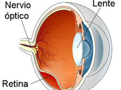
Retina is the innermost layer of the eye and is light sensitive in nature.
What is the Retina?
Retina is the innermost layer of the eye and is light sensitive in nature. When we see an object, the light rays pass through the lens in our eyes and fall on the retina. They get converted into neural signals/impulses here and the optic nerve carries these visual stimuli to the brain that translates them back as images. Now if you are a Harry Potter fan, then consider the retina as the platform 9 ¾ (the entry point to the world of magic). If something goes wrong here, then nothing reaches your center for imagination (the brain) and your vision to the beautiful world stays completely cut-off.

Diabetic Retinopathy
Diabetic retinopathy is one of the mostcommonly associated findings in diabetic patients. Diabetes affects the small blood vessels ofthe retina and weakens them, leading to bleeding in the eye, swelling of theretina, increased eye pressure and many other ways. Gradually it leads toblurring of vision, which if left untreated may lead to permanent vision loss.A lot of patients do not have any vision problem in the beginning, even thoughthe diabetic retinopathy may be very severe. This is the ideal window oftreatment where the vision is not greatly compromised and early prompttreatment can be more gratifying for the patient. Gradually it leads toblurring of vision, which if left untreated may lead to permanent vision loss.
NORMAL RETINA:

Diabetic retinopathy and associated visionloss:
The two main reasons for vision loss inDiabetic Retinopathy.
1. Diabetic Macular Edema:

Macula is the most important part ofretina for vision. In diabetic patients, retinal blood vessels are damaged andthe blood and fluid starts leaking and swelling gets collected at macula. Thisleads to blurring of vision.
2. Proliferative DiabeticRetinopathy:

In advanced Diabetes cases, theretina suffers from lack of oxygen. This leads to formation of new bloodvessels and bleeding over the retina, including macula.
VEIN OCCLUSION
Like any other tissue, retina is dependent on blood supplyfor its nutrients. The blood rich in nutrients comes through the retinalarteries, gets distributed into the capillaries and reaches the retina. Thenutrients are used by the retina and the waste products of metabolism isdistributed to the retinal veins and taken out of the eye.

In vein occlusions, the outflow passage is blocked partiallyor completely. When that happens, due to the backflow changes, the pressure inthe veins increases and leads to leaking of blood and fluid from the blockedveins and into the retina. If this involves the macula, there can be swellingof the macula and vision is affected.
Risk factors for retinal Vein Occlusion:
1. Diabetes
2. High Blood Pressure (Hypertension)
3. Overweight
4. Stress
5. High Cholesterols levels.
6. Cardiac patients.
Symptoms:
Normally, one eye is affected.
Treatment:
The treatment has to focus on the eye as well as thesystemic cause that lead to the vein occlusion.
1. Topical drops.
This can be line of management if there isno/minimal involvement of macula and the vision is not much affected. However,routine follow-up and OCT scan should be done to rule out macular involvementduring follow-up visits. Simultaneous physician and if required, cardiologistopinion might be advised by the retina specialist, if deemed necessary.
2. Intra-vitreal injections.
In cases that have significant macularedema affecting the vision, intravitreal anti-VEGF injections are thefirst-line of management. With regular treatment and follow-up, the macularedema subsides and vision is significantly restored.
3. Laser treatment.
In some patients, later on duringthe course of the disease, abnormal, new, immature blood vessels start to growbecause of the vein occlusion. When this happens, it might be necessary to uselaser treatment to arrest the growth of these new vessels and avoid bleedinginside the eye and further complications.
AGE RELATED MACULAR DEGENERATION (ARMD):
Macula is the central and the most visually important areaof your retina. With increasing age, the macula gets affected due to variousreasons. In elderly patients, the macula is likely to get more degenerated andhence the vision is affected. With ARMD, central vision is more affectedwhereas peripheral vision (i.e. Side vision) is not affected.
This is how a patuient with macular degeneration would seeas opposed to a normal person.
Types of ARMD:
There are two types of ARMD:
1. Dry ARMD:
This is the most common type (80%) out of the two and also lessvision-threatening compared to wet ARMD.With advancing age, the degenerated proteins get collected at the macula andlead to Dry ARMD.
2. Wet ARMD:
Insome patients(20%), dry ARMD if left untreated, might progress to wet ARMDstage. In this stage, new immature blood vessels start growing under themacula. As these vessels are immature, then tend to leak and sometimes bleedunder or inside the macula. This leads to a profound vision loss which ,if leftuntreated, can become permanent.
Many people don’t realizethey have ARMD until their vision is very blurry. This is why it is importantto have regular visits to a retina specialist. He/she can look for early signsof ARMD before you have any vision problems.
What are the risk factorsfor ARMD?
1. Age >50 years.
2. Smoking
3. High blood pressure (hypertensive patients).
4. Family history of ARMD
5. Overweight
6. Patients who tend to consume a high fat diet.
7. High blood cholesterol levels.
8. Over-exposure to sunlight (e.g. farmers,labourers etc.)
Symptoms:
- Difficulty in recognizing faces.
- Reduced clarity of central vision
- Need for brighter light while reading newspaper doing any near work
- Distorted vision
Treatment:
There is no cure forMacular Degeneration. However, vision loss can be slowed or stopped. Early treatment is important to control the disease before too much vision is lost
Dry ARMD patients have beenfound to benefit from taking a combination of certain nutritional supplementswhich may slow down the progression of ARMD. This was found in a large studiescalled AREDS and AREDS 2. Eye-healthy foods like Dark green leafy vegetables,yellow fruits and a balanced, nutrient-rich diet have been shown beneficial forpeople with ARMD. Food items rich in vitamins such as spinach, tomato, carrots,oranges etc might be consumed too.
Wet ARMD patients need activeintervention and treatment. In these patients, the mainstay of treatment isanti-VEGF injections. This helps to reduce the growth of new immature bloodvessels and reduces the fluid and blood leakage from them. Such patients mightneed multiple injections and need regular follow up with the retina specialist.
Home Monitoring with Amsler’sChart is very essential to look for progression.
How to check your vision athome with Amsler’s Chart:
- Keep the Amsler grid in a place where you see it every day. Many people keep an Amsler grid on their refrigerator door or on their bathroom mirror.
- In good light, look at the grid from about 12–15 inches away. Be sure to wear your reading glasses if you normally use them.
- Cover one eye. Look directly at the dot in the center of the grid with your uncovered eye. Notice if any of the lines look bent or wavy. See if any part of the grid looks blurry, dim, or out of shape.
- Now cover your other eye and test your vision this same way again.